Mrs. Z, a sixty years old lady with otherwise normal ealth had pain in her abdomen followed by vomiting. She went for an ultrasound examination of the abdomen, which revealed multiple gallstones and inflammation of gall bladder. In addition to this, she was found to have tumours in both her idneys. She underwent CT scan and then MRI, which confirmed tumours in both kidneys. This was devastating for the whole family. The panel of radiologists opined that these tumours could be cancerous in nature and must not be allowed to stay in the body. Usually nature gives paired organs, so that if there is injury or disease in one (kidney), then the other is sufficient to carry out function and support the life of individual. In this case both the kidneys had tumours, each one having potential to be fatal individually. The traditional standard treatment of such tumours is usually removal of the whole kidney. But thanks to Robotic technology, we planned to remove the tumours and save the kidneys. After detailed discussion with the family members and careful evaluation of the patient it was decided to remove the tumour from the left kidney first. We also decided to remove the gall bladder, which was the cause of pain to the patient. After laparoscopic removal of the gall bladder, the robotic surgery for left kidney tumor was started. Using Robotic technology, six small keyholes were made into the abdomen and the kidney was isolated from surrounding structures . The main blood supply of the left kidney, the artery and the vein were carefully cleared of all the surrounding fat and other tissues. These blood vessels were clamped and the blood supply to the kidney was stopped temporarily. Stop watch was started as we had twenty minutes to keep the blood vessels clamped, because taking longer time could be detrimental for the recovery of the kidney. The tumour was carved out using a sharp instrument and the cut margin of the kidney was stitched using special suture material. Having repaired the cut surface of the kidney, the clamps from the main blood vessels of the kidney were removed. Every one in the operation theatre was excited to look at the outcome. Thank fully the repair was perfect and there was no bleeding after removal of the clamps. We could manage to achieve all this in little less than twenty minutes. The patient was shifted to the ward after two hours and the next day she was made to take liquids orally. She was discharged on the third postoperative day. After five weeks, the same procedure was carried out on the right side. The patient withstood the procedure well and this time again she was discharged after three days. The kidney function assessed at the end of the second surgery revealed normal values.
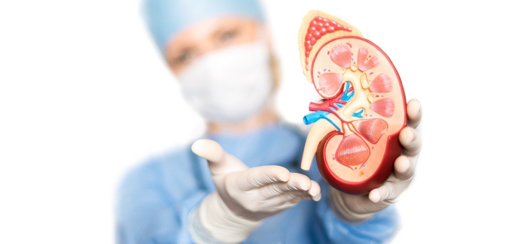
This is an example of appropriate use of state of the art technology to remove deadly tumours of the kidney while saving useful functioning kidney tissue. This is technically called Robotic Assisted Partial Nephrectomy. The risks. of the procedure has to be explained to the family and patient. There could be bleeding while isolating the blood supply of the kidney, thus forcing the surgeon to remove the whole kidney. While carving out the tumour, some tumour tissue might be left behind. After repair of the cut surface of the kidney, there could be cracking of kidney and bleeding, again forcing the complete removal of kidney. Occasionally, there might be leakage of urine from the cut surface of kidney, thereby necessitating a second operation to remove the leaking remaining kidney. However with the experience in such procedures, it becomes the standard of care for patients with kidney tumours.


