Public Sphere
Publications

Delivered a Plenary Lecture at 41 Annual conference of Society International de Urology
The Week
MedCrave
14 November, 2021, Dubai
November 29, 2020
July 30, 2020
The Hindu
April 09, 2016
Times of India
February 20, 2012
Hindustan Times
March 09, 2011
The Statesman
September 20, 2006
Delhi Times
January 22, 2006
Sahara Time Paranoma
August 27, 2005
Al-Yaum Daily
February 25, 2004
Qaumi Awaz
February 25, 2004
Daily Pratap Delhi
February 24, 2004
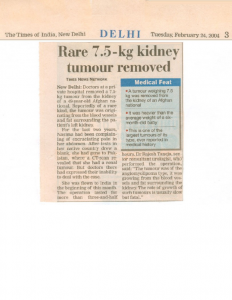
Times of India
February 24, 2004
The Indian Express
February 7, 2004
Rashtriya Sahara
September 18, 2003
Navbharat Times
April 30, 2003
Dainik Jagran
April 30, 2003
Punjab Kesari
Mustaqbil
Plus
January 21, 2006
Plus
April 23, 2005
The Pioneer
August 19, 2009
The Pioneer
December 10, 2008
The Pioneer
June 18, 2003
Articles
- Mail Today ePaper | July 06, 2010
- Uday India | June 19, 2010
- Times Life | December 25, 2005
- India Today | March 15, 2004
- Guardian Health Chronicle
- August 13 – August 16, 2009
- July 2 – July 29, 2009
- June 22 – July 5, 2006
- News Of The Week
- February 12 – February 18, 2005
- September 25 – October 01, 2004
This is the story of a 49-year-old lady, a native of Afghanistan. She had been suffering from bloating sensation in the abdomen and a steadily increasing abdominal girth for about three years prior to been seen by me. Of and on she would also experience dull ache in the left flank. She was investigated elsewhere and found to be having a giant tumour in the abdomen. With this information I saw her at a tertiary care hospital in New Delhi, India. A review of CT scan films revealed that a large fat containing tumour had replaced the left kidney while displacing the rest of the abdominal contents towards the other side of midline. The lady had been told earlier that this was an inoperable tumour. I decided to accept the challenging task and operated upon her on 7th February 2004 at Pushpawati Singhania Research Institute for liver Renal and Digestive diseases, New Delhi. The operation was conducted under general anaesthesia and lasted for three hours and twenty minutes.
A case report of a diabetic male who suffered from gas gangrene following urethral instrumentation underwent reconstructive procedure using local tissue flap. A 40 years old diabetic man underwent cystolithotripsy for a vesical calculus in a suburban nursing home. After removal of catheter he failed to pass urine and needed an indwelling catheter. Reinsertion of catheter could be possible only after the dilatation of urethra using a metal bougie. The patient developed fever and swelling of the external genitalia. At this stage he was referred to tertiary care center. On arrival in the emergency ward, he had clinical features of septicemia; diffuse swelling of external genitalia extending up to the lower abdomen. Subcutaneous crepitus was appreciated over the lower abdomen. A Computerized Tomography scan confirmed gas in the subcutaneous space, corpora cavernosal and corpus spongeosum. He was treated with broad-spectrum antibiotics, debridement and suprapubic diversion of urine. At a later date the left corpus cavernosum was excised and urethra was laid open. Once the sepsis was controlled, penis was reconstructed using a rotation flap using scrotal skin based on dartos fascia.
This is an unfortunate story of a woman whose life became miserable due to the most basic privilege of any lady of her age. While she was in labour during child birth, she did not receive adequate medical help resulting in what is technically called ‘Obstructed Labour’. In simple words, it means that the head of the unborn fetus gets stuck in the bony pelvis of the mother. As a result of this, the tissues of bladder get squeezed between the head of the fetus and the bone of the pelvis leading to ‘necrosis’ or breakdown of tissues. Consequent to obstructed labour, Mrs. S developed a hole in her urinary bladder (Vesico vaginal Fistula, or VVF) and started leaking urine continuously. She underwent three operations to close this hole in Gujarat, but unfortunately she continued to leak urine causing unabated physical and mental agony.
When Mrs. S came to PSRI hospital, she was diagnosed to have what is technically termed a persistent VVF. After examination and suitable investigations, Dr. Rajesh Taneja, senior consultant Urologist decided to take up the challenge. Mrs. S underwent a tedious abdominal surgery, made difficult due to previous operations, and lasting almost 6 hours. But at last, this was the end of miseries for Mrs. S and her family.
This is the story of a 19 year old boy S.D., who suffered liver failure at the age of 12 years. He underwent liver transplantation in 2002. Except for some initial problems after the transplant he was doing reasonably well till few months ago when he developed fever and jaundice again. He was found to have developed obstruction to the flow of bile due to formation of stones in the ducts within his liver. The ducts within his liver had become congested with pent up bile.
He underwent a procedure for draining the obstructed bile, which was done by passing a thin tube from the skin into the liver under ultrasound guidance. This relieved him of his fever and jaundice, but he continued to carry a tube and a drainage bag attached to his tummy. The surgical removal of stones and correction of the obstruction was considered and discussed with the parents of the child. In view of the high risk of this operation including fatal complications, his parents did not give consent for the surgery.
